Erbe Vision: Improving imaging systems for minimally invasive surgery
Erbe Vision’s Chief Executive Officer Shirish Joshi is an expert in minimally invasive surgery imaging systems. In this interview, he talks about the potential of fluorescence imaging technologies, augmented reality as an enabler for the surgery of the future, and why innovations in endoscopy need the best light.
Erbe Vision GmbH
What is Erbe Vision's approach to improving surgery?
We asked ourselves what enables better surgery and decided that our answer is surgical precision. We work in the field of minimally invasive surgery, which offers many benefits over conventional methods like open surgery, but also has clear limitations. For example, the surgeon can no longer rely on haptic sensations to help diagnose or treat unhealthy tissue. The use of tools deprives the surgeon of this tactile sensation, especially as more and more surgeries are being performed with the help of robots. The loss of one of the senses has to be replaced with other information, especially visual information.
Please give some examples.
Surgeons have become more dependent on the color of tissue to make decisions. Everything appears in various shades of red, making fine color gradations ever so important. As the surgeons have to navigate by sight, inaccuracy in colors and images can lead them to wrong judgements and wrong tissue cuts before performing the “right” ones. The goal of our company is to provide surgeons with the best information possible to enable them to work as precisely as possible. Hence, we bring a new dimension to surgical precision by developing improved imaging technologies.
The field of imaging has evolved strongly in recent times. New generations of monitors are able to show one billion colors versus some 17 million colors before. Near-infrared fluorescence imaging provides us with much more additional information. These developments help us to enable better surgery.
What role does light play in the visible and non-visible spectrum?
Light is the most important part of the imaging system! We all know this from photography where light is more important than the camera itself: Better light creates better imaging. 99% of the photographer’s work goes into arranging the light, and then the actual photograph is taken in one click. In surgery, we don’t have this luxury. We need to get the right type of light inside the body, and color accuracy very much depends on the kind of wavelengths used. To a layman’s eye, various tissues look similar, but during surgery, the fine gradations of light become relevant to reveal their different shapes of red.
The whole optical system is important: excellent light sources, filters, light guides, endoscopes etc. are needed. Light has a large impact on the type of imaging we are able to generate. And the imaging itself has a very high importance in minimally invasive surgery. We are working mostly on fluorescence imaging in the near-infrared (NIR) wavelengths, which do not disturb the natural images in the visible spectrum. Thus, we aim to provide live NIR imaging without any quality degradation of regular white light imaging.
How will Erbe Vision's innovative fluorescence-guided surgery system support surgical procedures?
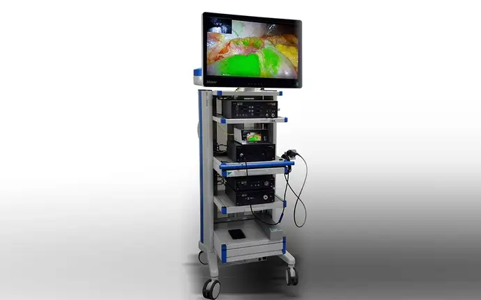
We were the first company in the world to introduce 4-chip cameras for fluorescence-guided real-time surgeries without compromising visible light imaging. The additional image information provided by fluorescence allows the surgeon to visualize critical structures such as bile ducts, liver segments, sentinel lymph nodes, tissue perfusion, and blood supply in a better way. What we do here is to inject a dye called indocyanine green (ICG) into the patient. This fluorescent dye functions as a contrast agent which allows us to see critical structures better. This is one step in fulfilling our mission of improving surgical precision, which would not be possible without these new technologies.
Please explain your work around augmented surgery.
We partnered with Ludwig-Maximilians-University (LMU) Clinic in Munich as well as the Technical University of Munich (TUM) to develop a navigation system for minimally invasive surgery. We want to bring augmented reality-assisted surgery to the operating room. We have been working on this project “ATLAS” for four years already. The basic architecture and the first prototypes are ready, and we also completed animal tests to determine reallife accuracy. The project was funded with 1.9 million Euros by the German Federal Ministry of Education and Research.
What exactly are you working on within this project?
The success of minimally invasive surgery depends on the quality of the information available to the surgeon. Sometimes even the best lighting and imaging doesn’t allow surgeons to see where they are navigating in the body, especially when too much blood is involved. Surgeons use pre-operative CAT and MRT images from different angles which they must memorize and then match with what they see live during surgery. It takes a lot of training and experience to navigate within the abdomen, and it is virtually impossible to be perfectly accurate when working like this. The demands on the skills of the surgeons are extremely high.
What we aim to do is aid the surgeons in this process. We create a 3D map of the CAT of the patient, segment critical structures, and overlay them on the live laparoscopic image in real time. This gives the surgeon as much context-specific information as possible to navigate very precisely within a patient’s body, and to be very aware of the area they are working in. Our system will provide this important guidance.
The success of Erbe Vision is based on the concept of including customer feedback in its development activities. How does this allow you to offer better products to the market?
We work very closely with end-users, i.e. the surgeons, starting at the conceptualization stage. As we are working on several break-through technologies such as fluorescence-guided surgery or surgical navigation systems, it is important to identify clinical needs so that our products can really fulfill the requirements of the surgeon and finally be accepted on the market.
Where do you see endoscopy in 10 years' time?
We believe that not only endoscopy, but the whole operating room will become more and more digital. Surgeons will have more information at their disposal to make better decisions. They will have better tools to perform more precise surgery, and many surgeries will use robots because we are short of manpower, especially highly-qualified experts such as surgeons and operating room nurses. The robots help the surgeons to operate more precisely and in more ergonomic positions. Therefore, surgeons will be able to work longer both during the day, but also during their career.
In the end, these developments might even necessitate the redesign of the operating rooms and a re-training of the operating room staff. The increase in productivity should be able to offset the higher costs. For simple procedures, we expect more and more single-use endoscopes to be used.
Text: Dr. Haike Frank, SCHOTT
All photos: Erbe Vision
January 6, 2021; updated September 8, 2022
Links