TracInnovations: Delivering patient motion correction to improve image-based diagnostics

Why is motion tracking in MRI procedures a promising field to work in and how do you help to advance medical technology?
MRI scanners represent an increasing market, and there is a growing number of MRI scanners, not only in developed countries, but all over the world. As it is still expensive equipment, we see our primary market in North America and Europe, but we also see potential in the rest of the world.
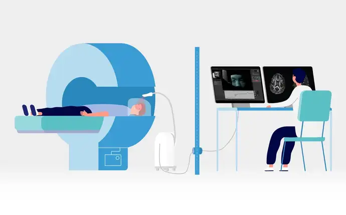
Using MRI technology is the safest way to perform a 3D scan of a patient, as it does not use radiation needed in other traditional scan methods such as X-ray, PET or CT. However, there is one restriction to MRI technology: Unwanted patient motion degrades the image quality. When patients go through an MRI examination, they are usually required to stay still for about 40 minutes. During that time, the scanner will take approximately 5 to 10 images, so each image can take several minutes to record. If the patient moves just a few millimeters during that time span, the quality of the images will decrease considerably.
Many studies have been conducted on motion and the problem of patient motion during a scan. Motion during MRI results in the need for repeated sequences in about 20% of all scans*, and as scanners can be very expensive and run up to 24/7, there is a huge potential for improvement. This study estimates an indirect cost to the hospital of €97,000 per scanner per year due to motion artifacts. Another research paper estimates that children and patients with involuntary head motion are often not considered for scanning or scanned with sedation resulting in costs of approx. €260,000 per scanner per year**.
With our solution, we address this problem of patient motion leading to corrupted images. We can actually accept that a patient moves during the scan because our system will monitor and record the motion information and feed it back to the scanner in real time, enabling it to adjust according to the movement (research only). In addition, we can also reduce the need for sedation that is commonly used among young children.
Please introduce your innovation Tracoline, a Markerless Motion Tracking System.
Our motion tracking system – an add-on for the MRI scanner –, monitors and tracks the motion of patients during an MRI scan. We can use this information to alert the personnel that the person is moving too much, or we can even use it to feed the motion information back to the scanner so that the scanner can adjust and create a more accurate and better quality 3D image of the patient.
Our Tracoline system is located behind the scanner. We use glass optical fibers to extend the vision of the camera from its location on the trolley into the scanner, allowing us to look directly at the patient. There is a very strong magnetic field inside the scanner into which we cannot place electronics. The optical fibers which are interference-free extend from the backside of the scanner all the way through the scanner bore to the vision probe located just on top of the patient’s head.
At TracInnovations, we produce both the hardware system consisting of the camera and trolley, as well as the TracSuite software package, which has a simple-to-use graphical user interface. It captures 3D surfaces of the object that it is looking at and uses this information to continuously track the motion of the patient. A video monitor provides a real time patient image and a digital output continuously of any movement in all six degrees of freedom.
Tracoline is an advanced 3D stereo vision system which monitors and captures movements online. It is developed to fulfill the clinical requirements for a user-friendly setup, reliable tracking quality and real-time motion feedback. The markerless system is invisible to the patient and requires minimal additional work from the scan staff.
Tracoline constructs high quality 3D surfaces from structured invisible IR light, enabling markerless tracking of the patient’s head – all supporting the operational workflow and patient comfort. Tracoline is delivered with a TracSuite software package focusing on robust tracking. It is simple to use with a Graphical User Interface for real-time monitoring of patient movement.
How do you “upgrade” from motion tracking to motion correction?
In order to perform motion correction, you need a tracking device, which provides motion tracking data. Optimally, the motion correction then happens on the scanner itself in real time. This means that the scanner vendors also have to update their scanner software in order to use the information sent by the motion tracking system. We cooperate very closely with research sites because they can develop this kind of software that runs on their scanners. Together with the research teams, we cover the whole pipeline of motion tracking and correction to improve the quality of the images.
Our solution is scanner-independent which means that it is compatible with MRI scanners from the major scanner vendors Siemens, GE, and Philips. We are trying to push the vendors to open their software interfaces and release software that is already applicable to our motion tracking system. We think that will come in the near future.
Right now, that entire pipeline of motion tracking and correction can only happen in a research environment. Our motion tracking system alone can be used on clinical patients, but we would currently only give the information back to the scanner operator so that they can make a judgement whether to rescan the patient if they see too much motion. We are convinced that – together with the scanner vendors – we can leverage the full potential of our innovation and enable motion correction as part of the MRI scan process.
What is the status of the market launch of your system?
We were FDA cleared in April 2021, so we can now market our motion tracking system in the USA to the clinical market. We are very proud of this.
We are currently also working on receiving CE marking for Europe and expect to be successful in the near future. Due to the challenges of the MDR regulation, the process has been taking a bit longer.
Why do you use infrared light, and how does this improve the patient’s experience as well as the quality of the images?
We have a stereo vision system that is based on the combination of an IR camera and a projector. That gives us stereo capabilities to create a 3D image of the patient. We use a technique called structured light where we use the projector to send a specific pattern onto the surface of the patient, and then record the same image with the camera. We are using IR wavelengths because they are non-visible to the human eye so the patient will never notice that motion tracking is being performed. This is an important feature, which makes us unique compared to competing technologies needing markers attached to the patient’s face.
The technology would also work with visible light, but it would be annoying for the patients to see the light flashed into their face. We do not only improve patient comfort, which is an important feature for medical devices, we also support the operational workflow by improving tracking accuracy by tracking the patients directly, not markers attached to their faces, thus reducing one layer of complexity. The workflow is simple and automated and requires no additional scan time or patient preparation.
What does your cooperation with renowned hospitals look like?
Our developments and innovations strongly rely on cooperation with research institutions. We typically work with the research institutions of larger hospitals, for example in Boston, Copenhagen and Stockholm***. Here, we can establish a strong connection between our technology, the research done on volunteers who test our new implementations, and finally the clinical parts of the hospital where we are currently running clinical trials.
On a more personal note: as a computer scientist, what inspires you about working for a tech-start-up?
It has always inspired me to see how computer science can be used to advance technology. To work on something cutting edge is really interesting. In a smaller start-up company like TracInnovations, there is such a short distance from our work to the benefit of the patient that I can immediately see our efforts going into a product that will be used tomorrow. All of us in TracInnovations work with a common goal and we are all very passionate to have this succeed.
I started here in 2016, was hired by the three founders as the first employee, and given the responsibility to improve the software part of the product. My background is in high-performance computing, so I was very happy to work on a project around computer vision to solve a real and serious problem to the benefit of patients. Computer vision, artificial intelligence and machine learning are very interesting scientific fields to work in. I am proud to say: we do a little bit of good in the world every day.
Text: Dr. Haike Frank, SCHOTT
All photos: TracInnovations
*Jalal B. Andre et al., JACR 2015
** Jakob Slipsager et al., JMRI 2020
*** Boston Childrens’ Hospital (USA), Athinoula A. Martinos Center for Biomedical Imaging, at Massachusetts General Hospital, Boston (USA), Rigshospitalet, Copenhagen (Denmark), Karolinska Institutet, Stockholm (Sweden)
July 14th, 2021